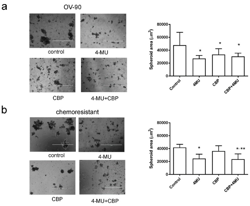
Figure 4.
Effects of 4-methylubelliferone (4-MU) on spheroid formation. Representative images of spheroids formed by (a) OV-90 and (b) one of the chemoresistant primary ovarian cancer cells treated with control, 4-MU (1 mM), carboplatin (CBP, 100 µM), and 4-MU (1 mM) + CBP (100 µM) for 72 h. Data are expressed as median area of spheroids >200 µm in diameter (95% confidence interval, CI, n = 34–106) in five fields/treatment groups from 3–4 independent experiments. *, significantly different from control, **, significantly different from CBP treatment (p < 0.05, Kruskal–Wallis test, Dunn’s multiple comparison test). Scale bar = 1000 µm.

