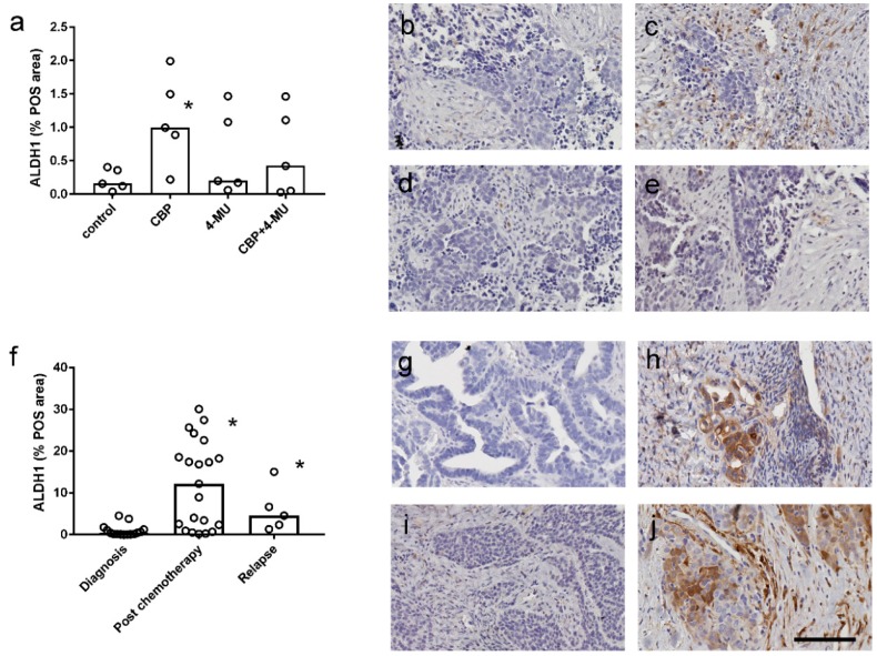
Figure 6.
Effect of chemotherapy and 4-methylubelliferone (4-MU) on ALDH1 immunostaining in ovarian cancer tissues. (a) Quantitation of ALDH1 immunostaining in serous ovarian cancer explants tissues (n = 5) treated with control media, carboplatin alone (100 µM), 4-MU alone (1 mM), and CBP + 4-MU in combination. Data are expressed as percentage positive area (% POS) measured by video image analysis. *, Significantly different from control treatment (p = 0.0087, Friedman test, Dunn’s multiple comparison test). Representative images of ALDH1 immunostaining in the explant tissues treated with control media (b), CBP (c), 4-MU (d), and CBP+4-MU (e). (f) Quantitation of ALDH1 immunostaining in serous ovarian cancer tissues at diagnosis (n = 15), following neoadjuvant chemotherapy (n = 20) and at recurrence (n = 5). Data are expressed as % POS area. *, Significantly reduced from level at diagnosis (p = 0.0002, Kruskal–Wallis test, Dunn’s multiple comparison test). ALDH1 immunostaining in matched patient tissues at diagnosis (g) and following neoadjuvant chemotherapy (h). ALDH1 immunostaining in matched patient tissues at diagnosis (i) and at relapse with chemoresistant disease (j). All images are the same magnification. Scale bar = 100 µm.

