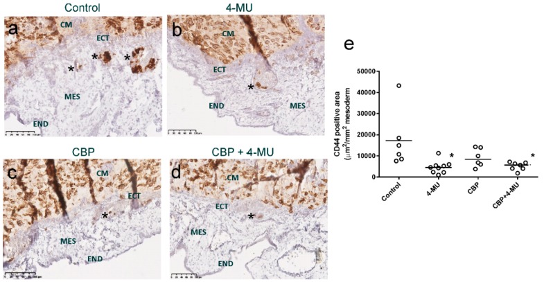
Figure 7.
4-MU and carboplatin treatment inhibit in vivo invasion of chemoresistant primary cells. Representative images of the invasion of chemoresistant primary ovarian cancer cells shown by CD44 immunostaining. Treatment includes (a) control media, (b) 4-MU (1 mM), (c) carboplatin (CBP 100 µM, and (d) CBP 100µM + 4-MU (1 mM). Paraffin sections were immunostained with a CD44 mouse monoclonal antibody. Asterisks show examples of CD44 positive cancer cells that have invaded into the mesoderm (MES). (e) Quantitation of chorioallantoic membrane (CAM) invasion into the mesoderm. Data are of the CD44-positive area (µm2/mm2 of mesoderm) from 5–9 chick embryos implanted with chemoresistant primary cells from one patient. *, Significantly different from control (p = 0.0272, Kruskal–Wallis test, Dunn’s multiple comparison test). CM = cancer cells in matrigel implant, ECT = ectoderm, MES = mesoderm, END = endoderm. Scale bar = 100 µm.

