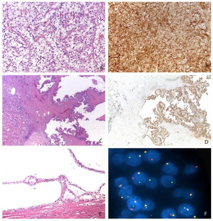
Figure 2.
Different morphologies of Xp11 translocation renal cell carcinomas: resembling a clear cell renal cell carcinoma (Magnification: 200×) (A), showing a papillary (Magnification: 25×) (C) or cystic (Magnification: 100×) (E) pattern. An example of strong and diffuse expression of cathepsin K (Magnification: 200×) (B), the nuclear positivity of PAX8 (Magnification: 25×) (D), and the demonstration of TFE3 gene rearrangement by FISH (Magnification: 1000×) (F).

