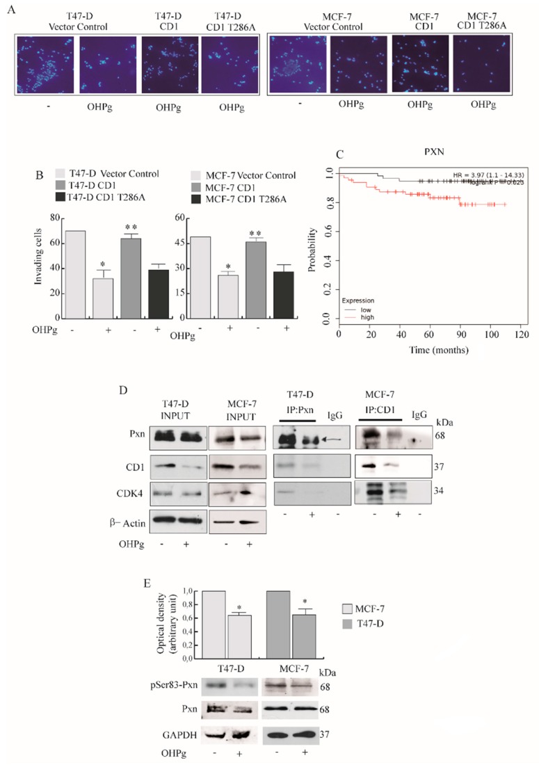
Figure 5.
OHPg effects on CD1/Cdk4/Paxillin (Pxn) interaction and Pxn phosphorylation. (A) Transmigration assay, (B) Invasion assay. Cells were co-transfected with vector control, CD1 or phosphorylation site mutant of CD1 (CD1 T286A) expression plasmids. Columns are the mean of three independent experiments each in triplicate; bars, SD; * p ≤ 0.05 vs. vehicle treated vector control cells. ** p ≤ 0.05 vs. OHPg-treated vector control cells (C) Kaplan–Meier distant metastasis-free survival analysis in luminal A PR+ breast carcinoma patients (n = 122) with high and low Pxn expression analyzed as described in the Materials and Methods. Kaplan-Meier survival graph, and hazard ratio (HR) with 95% confidence intervals and logrank p value (D) Co-immunoprecipitation analysis (right panel). Cytoplasmic extracts were immunoprecipitated with anti-Pxn or anti-CD1 antibodies, as indicated, and immunoblotted with anti-Pxn, anti-CD-1 and anti-Cdk4. Input (left panel), samples without immunoprecipitation. βActin, loading control. IgG was used as the negative control. (E) Immunoblotting for pSer83 Pxn and Pxn expression in T47-D and MCF-7 cells, as indicated. GAPDH, loading control. Columns indicate the mean of relative ratio pSer83 vs. total Pxn. * p ≤ 0.05 vs. vehicle-treated cells.

