Figure 2.
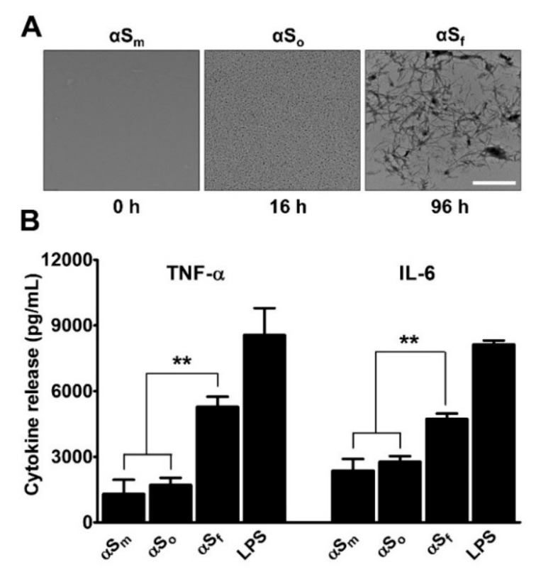
Impact of α-synuclein species on microglial cytokine release. (A) Transmission electron microscopy images of αS (70 µg/ml) samples agitated at 37 °C by orbital shaking and harvested after 16 h or 96 h. Images show the presence of oligomers (αSo) and fibrillary species (αSf), at 16 and 96 h, respectively. Scale bar: 1 µm. (B) Impact that samples of αS (70 µg/mL) harvested after 16 h (αSo) or 96 h (αSf) of orbital agitation and non-shaken samples (αSm) have on the release of tumor necrosis factor (TNF)-α and Interleukin (IL)-6 in microglial cell cultures. Cytokine release was near to zero in control cultures. Lipopolysaccharide (LPS) (10 ng/mL) was utilized as a positive control for stimulation. Data are means ± standard error of the mean (SEM) of three independent experiments performed in triplicate. Note: **p < 0.01 vs. αSf.

