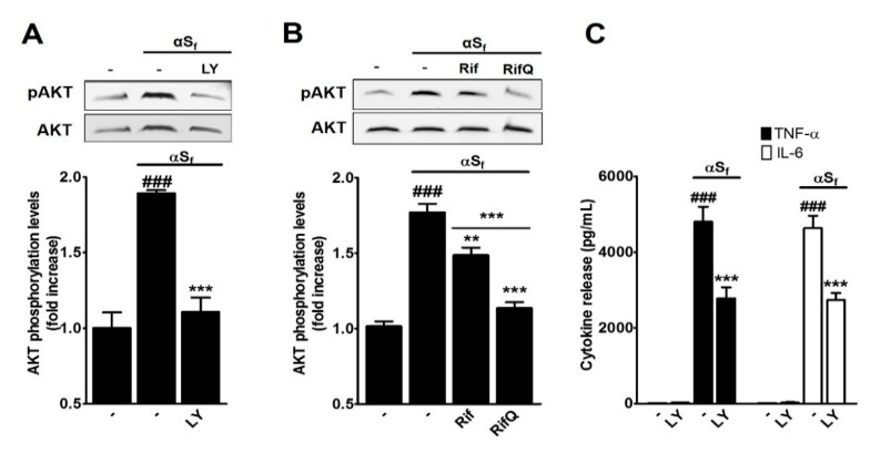
Figure 5.
Rif and RifQ inhibit a PI3K-dependent mechanism. (A) Western blot analysis of protein kinase B (AKT) phosphorylation in control and αSf-treated cultures exposed or not to the PI3K-specific inhibitor LY (2.5 μM). (B) Western blot analysis of AKT phosphorylation in control and αSf-activated microglial cells treated or not with 100 μM of Rif or RifQ. A representative blot is included (top), and relative levels of pAKT/AKT are shown (bottom). (C) TNF-α (filled bars) and IL-6 (open bars) released by microglial cells pretreated or not with LY prior to stimulation with αSf. Values are shown as means ± SEM of four independent experiments performed in triplicate. Note: ###p < 0.001 vs. controls. **p < 0.01 vs. αSf and ***p < 0.001 vs. αSf or Rif vs. RifQ.

