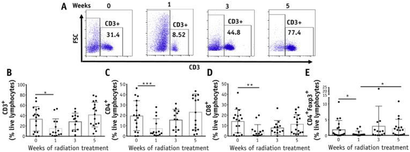
Fig. 1.
Changes in T-cell frequencies in the cervix throughout chemoradiation therapy (CRT). (A) Representative plots for CD3+ T cells in the cervix from 1 patient before and after 1, 3, and 5 weeks of treatment. (B) Percentages of CD3+, CD4+, CD8+ and CD4+FoxP3+ T-cell subsets among live lymphocytes were quantified (n = 20).

