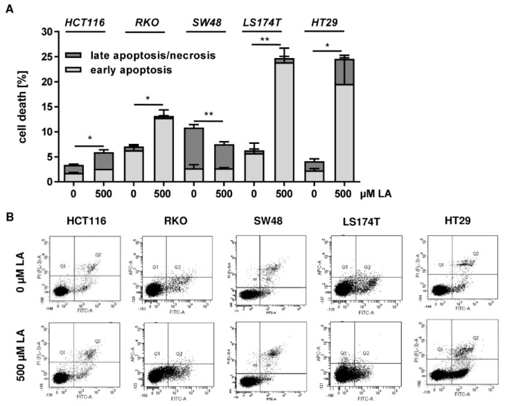
Figure A1.
Cytotoxicity of LA in different cell lines after 48 h. (A) Flow cytometric cell death measurement using AnnexinV/PI staining in a panel of CRC cells including HCT116, RKO, SW48, LS174T, and HT29. CRC cells were incubated with 500 µM LA or the solvent control EtOH (0 µM) for 48 h. Data is presented as mean + SEM (n ≥ 3). * p < 0.05; ** p < 0.01. (B) Representative dot plots of (A).

