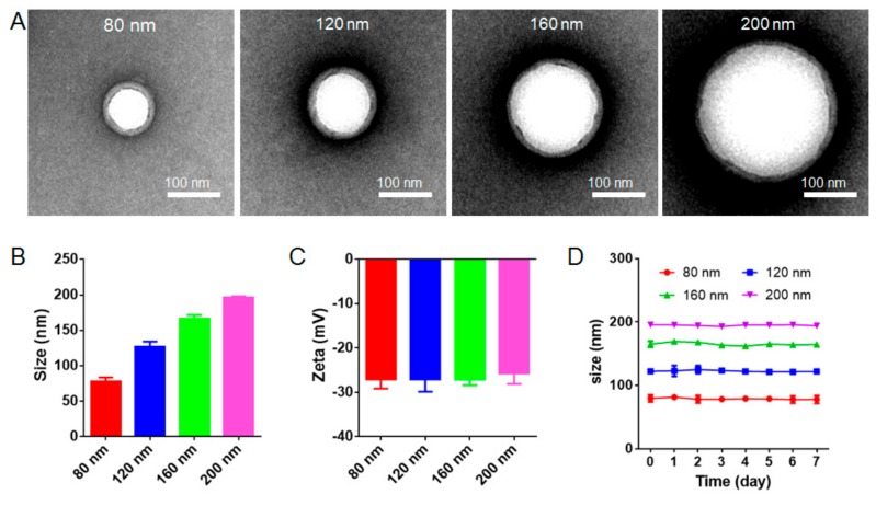
Figure 1.
Characterization of red blood cell membrane-coated nanoparticles (RBC-NPs) with different diameters. (A) Transmission electron microscopy (TEM) images of RBC-NPs with diameters of 80 nm, 120 nm, 160 nm, and 200 nm demonstrated the core/shell structure of RBC-NPs. Scale bar = 100 nm. (B) Size and (C) surface zeta potential of RBC-NPs with four different diameters (n = 3). (D) Diameter-dependent stability of different-sized RBC-NPs at 4 °C for 7 days (n = 3). All error bars represent standard error of the mean.

