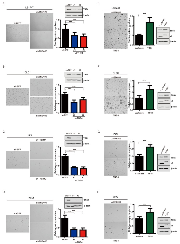
Figure 3.
TNS4 expression is associated with the oncogenic potential of colon cancer cells. (A–D) LS174T (A), DLD1 (B), DiFi (C) and WiDr (D) cells stably expressing either shTNS4 or shGFP were used for colony formation assay in soft agar. The relative number of colonies formed by shTNS4 bearing cells are depicted as bar graphs (n = 3, mean + SD) following normalization to that of shGFP-bearing cells. Cell lysates prepared from the same cells were subjected to immunoblotting analysis with antibodies against TNS4. Representative photomicrographs of colonies formed in soft agar are shown. (E–H) LS174T (E), DLD1 (F), DiFi (G) and WiDr (H) cells ectopically expressing either TNS4 or luciferase were used for colony formation assay in soft agar. The bar graph depicts the relative number of colonies formed by TNS4 expressing cells following normalization to that of cells expressing luciferase (n = 3, mean + SD). Representative photomicrographs of colonies formed in soft agar are shown. Cell lysates prepared from LS174T, DLD1, DiFi and WiDr cells were subjected to immunoblotting with antibodies against TNS4 and V5-tag.

