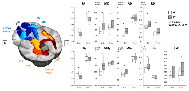
Figure 3.
(A) Region-of-interest analyses of the anterior (ACR, ATR, and forceps minor) and posterior (PCR, PTR, and forceps major) white matter areas. (B) Mean values for DTI (FA, MD, AD, and RD) and FW imaging (FAT, MDT, ADT, RDT, and FW) indices of the anterior and posterior white matter areas in healthy controls (white bars) and patients with PD (gray bars). ACR, anterior corona radiata; AD, axial diffusivity, ADT, FW-corrected axial diffusivity; ATR, anterior thalamic radiation; DTI, diffusion tensor imaging; FA, fractional anisotropy; FAT, free water-corrected fractional anisotropy; FW, free water; HC, healthy controls; MD, mean diffusivity; MDT, free water-corrected mean diffusivity; PCR, posterior corona radiata; PD, Parkinson’s disease; PTR, posterior thalamic radiation; RD, radial diffusivity; RDT, free water-corrected radial diffusivity.

