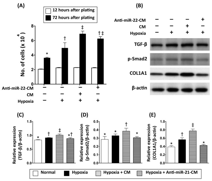
Figure 4.
Inhibition of miR-21 reduced the effect of conditioned medium on activation of cardiac fibroblasts. (A) Cell number counting to determine the proliferative rate of cardiac fibroblasts incubated with hypoxia (3% O2) and in conditioned medium derived from OGD-treated cardiomyocytes. (B) Western blots to examine the expression levels of fibrosis signaling proteins (TGF-β, phosphorylated Smad2) and collagen deposition (collagen typ1 1 alpha 1, COL1A1) in primary cardiac fibroblasts incubated with hypoxia and in conditioned medium derived from OGD-treated cardiomyocytes. (C) Quantitative data represent the ratio of TGF-β expression to β-actin expression. (D) Quantitative data represent the ratio of phosphorylated Smad2 to β-actin expression. (E) Quantitative data represent the ratio of COL1A1 to β-actin expression. CM, conditioned medium derived from OGD-treated cardiomyocytes. Anti-miR-21-CM, conditioned medium derived from cardiomyocytes pre-transfected with Anti-miR-21 (20 nM). Groups with different symbols (*, †, ‡), p < 0.05. n = 6 for each group.

