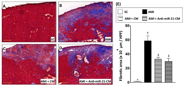
Figure 7.
Conditioned medium derived from cardiomyocytes with oxygen–glucose deprivation decreased myocardial collagen deposition in rats with myocardial infarction. (A) Histopathological examination on sham control myocardial section with Masson’s trichrome (MTC) staining. (B) MTC staining for myocardial section from rats with AMI induction. (C) MTC staining for myocardial section from rats with AMI induction and injection of conditioned medium derived from OGD-treated cardiomyocytes. (D) MTC staining for myocardial section from rats with AMI induction and injection of conditioned medium derived from OGD-treated cardiomyocytes with anti-miR-21 pre-transfection. (E) Calculation of collagen disposition area in myocardium. SC, sham control. AMI, acute myocardial infarction. CM, conditioned medium derived from OGD-treated cardiomyocytes. Anti-miR-21-CM, conditioned medium derived from cardiomyocytes pre-transfected with anti-miR-21. Groups with different symbols (*, †, ‡), p < 0.05. n = 8 for each group. Scale bar in left lower corner indicates 200 μm.

