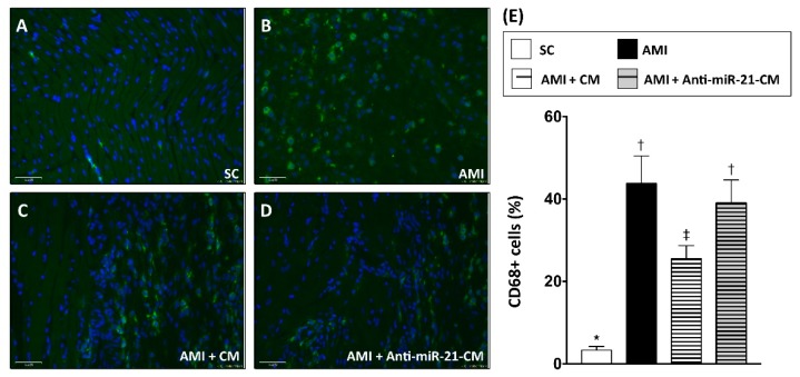
Figure 8.
Inhibition of miR-21 reduced the effect of conditioned medium on ameliorating myocardial infiltration of CD68+ macrophages. (A) Immunofluorescent staining to detect the distribution of CD68+ macrophages in sham control myocardial section. (B) Immunofluorescent staining to detect the distribution of CD68+ macrophages in myocardial section from rats with AMI induction. (C) Immunofluorescent staining to detect the distribution of CD68+ macrophages in myocardial section from rats with AMI induction and injection of conditioned medium derived from OGD-treated cardiomyocytes. (D) Immunofluorescent staining to detect the distribution of CD68+ macrophages in myocardial section from rats with AMI induction and injection of conditioned medium derived from OGD-treated cardiomyocytes with anti-miR-21 pre-transfection. (E) Calculation of CD68+ cells in myocardium. SC, sham control. AMI, acute myocardial infarction. CM, conditioned medium derived from OGD-treated cardiomyocyte. Anti-miR-21-CM, conditioned medium derived from cardiomyocyte pre-transfected with anti-miR-21 (20 nM). Groups with different symbols (*, †, ‡), p < 0.05. n = 8 for each group. Scale bar in left lower corner indicates 50 μm.

