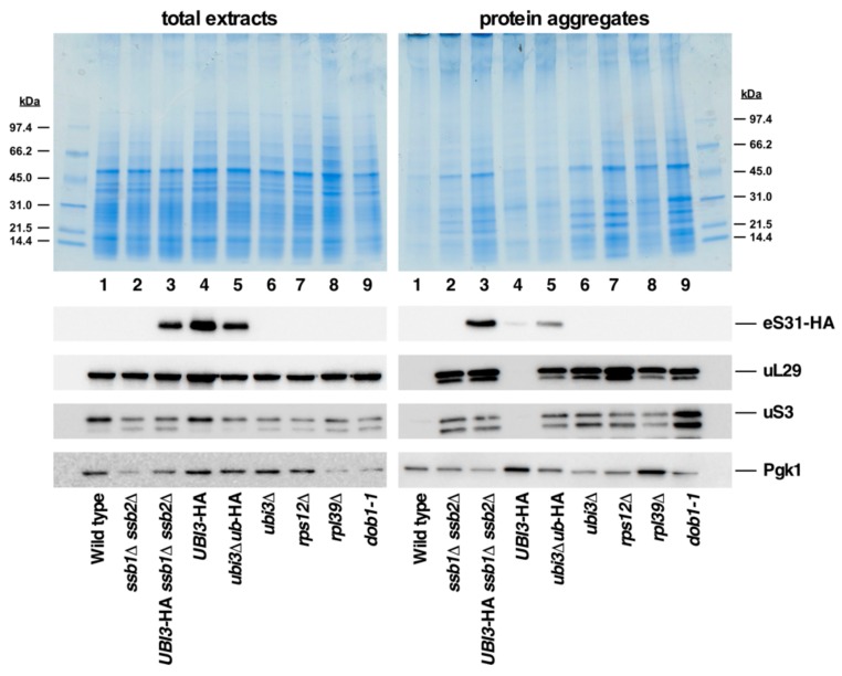
Figure 9.
Analysis of protein aggregation in the ubi3∆ub-HA strain and cells lacking individual r-proteins or being defective in ribosome biogenesis. Strains W303-1A (Wild type), JDY532 (ssb1∆ ssb2∆), SMY324 (UBI3-HA ssb1∆ ssb2∆), TLY56.D3 (UBI3-HA), TLY61.A2 (ubi3∆ub-HA), TLY14.3C (ubi3∆), SMY315 (rps12∆), ORY211 (rpl39∆), and MS157-1A (dob1-1) were grown to logarithmic phase in YPD medium at 30 °C. Then, total protein extracts and protein aggregates were prepared, separated by SDS-PAGE, and stained with Coomassie (upper part) or subjected to western blot analysis (lower part) using anti-HA (detection of eS31-HA), anti-uL29, anti-uS3, and anti-Pgk1 antibodies.

