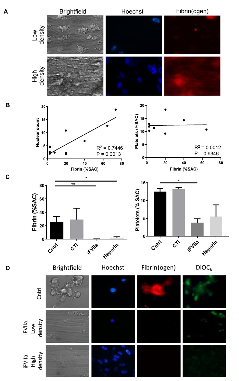
Figure 2.
Recalcified citrate anticoagulated whole blood was perfused over HHALPCs with cells seeded at either low or high density. Microscopic images were taken after 6 min of blood perfusion. Cells were stained with Hoechst (blue) to visualize cell nuclei, while Alexa fluor (AF)647-fibrinogen (red) and DiOC6 (green) were used to measure fibrin formation and platelet activation, respectively. Fibrin surface area coverage (SAC) was analyzed using Fiji software. (A,B) Note a positive linear correlation (R2 = 0.7446, p = 0.0013) between cell density and fibrin generation. (C,D) Fibrin generation and platelet activation was significantly reduced when inactive FVIIa (iFVIIa) (p < 0.01) was supplemented. On the contrary, corn trypsin inhibitor (CTI) did not interfere with fibrin generation or platelet activation. Data are presented as mean + SEM, non-parametric test (Kruskal–Wallis) * p < 0.05, ** p < 0.01.

