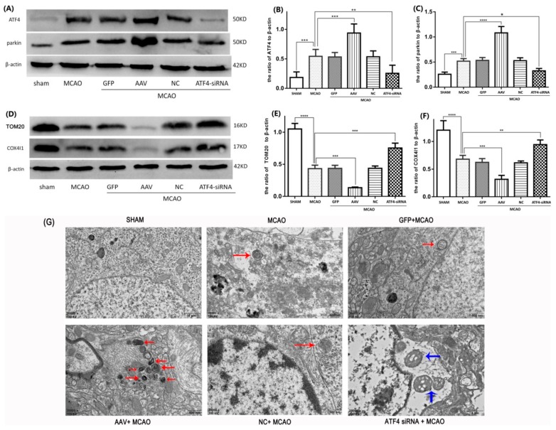
Figure 3.
ATF4 increases parkin expression and mitophagy activity during cerebral I/R injury in rats. (A–C) Representative Western blots of ATF4 and parkin. (D–F) Representative Western blots of TOM20 and COX4I1. Protein levels were normalized to β-actin. (G) The representative ultrastructures of mitophagy are shown: the structures of mitochondrials and nucleus were normal in the sham group; a few mitochondria engulfed by a double-membrane structure are labeled by red arrows in the MCAO, GFP + MCAO and NC + MCAO groups; many autophagic vesicles, which encapsulated with mitochondrias and were fused with lysosomal, were labeled by red arrows in the AAV + MCAO group; in the ATF4-siRNA + MCAO group, swollen mitochondrias were labeled by blue arrows and the typical autophagic vesicle was hardly observed. Error bars represent mean ± SD. (* p < 0.05, ** p < 0.01, *** p < 0.001 and **** p< 0.0001 vs. indicated group). n = 5 in each group. MCAO, middle cerebral artery occlusion-reperfusion; GFP, green fluorescent protein; AAV, ATF4 overexpression adeno-associated virus; siRNA, small interfering RNA; NC, negative control siRNA.

