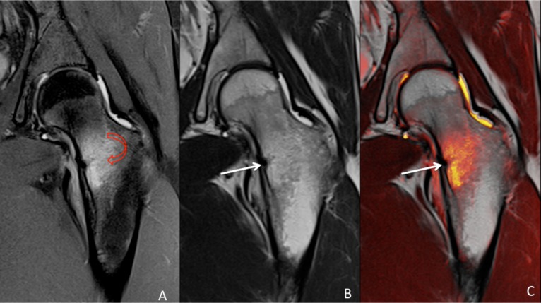
Figure 1.
MRI of the left hip. Coronal view showing minimal cortical hypointensity in the medial aspect of the femoral neck, indicating compression side fracture (white arrow), with extensive surrounding oedema (red arrow). (A) proton density–weighted image, (B) T1-weighted image and (C) fusion image.

