. 2019 Sep 3;8:e47615. doi: 10.7554/eLife.47615

Figure 1. Maximum likelihood phylogenetic tree based on MAFFT alignments of the predicted replicase protein of (A) Salmon pescarenavirus and related arenaviruses, (B) Chinook Aquareovirus and related Aqua and orthoreoviruses and (C) Pacific salmon nidovirus and related Nidovirales.

Sequences from this study are marked with an asterisk, scale bar represents the number of amino substitutions per site, node labels show the bootstrap support and host groups are shown by colour. Trees are mid-point rooted, so do not necessarily represent the ancestral relationship of the viruses. Amino acid alignments have been provided in the source data for Figure 1.

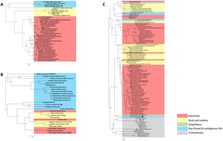
Figure 1—source data 1. Arenavirus amino acid alignment.
DOI: 10.7554/eLife.47615.006
Figure 1—source data 2. Nidovirus amino acid alignment.
DOI: 10.7554/eLife.47615.007
Figure 1—source data 3. Reovirus amino acid alignment.
DOI: 10.7554/eLife.47615.008
Figure 1—source data 4. Arenavirus phylogenetic tree.
DOI: 10.7554/eLife.47615.009
Figure 1—source data 5. Reovirus phylogenetic tree.
DOI: 10.7554/eLife.47615.010
Figure 1—source data 6. Nidovirus phylogenetic tree.
DOI: 10.7554/eLife.47615.011

Figure 1.

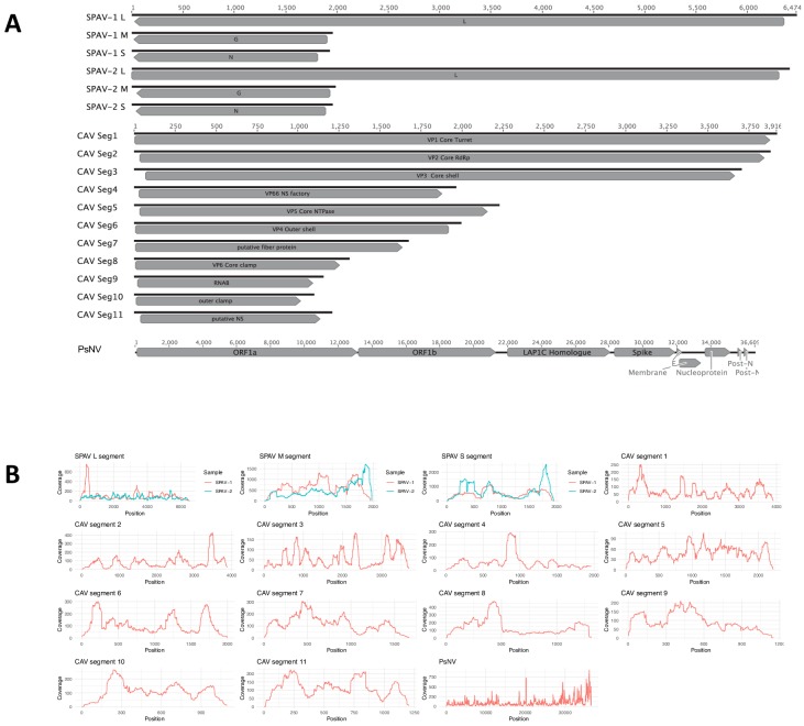
Figure 1—figure supplement 1. Genome organisation and coverage.

Figure 1—figure supplement 1.

(A) Genome organisation, ORF prediction and (B) Genome coverage for Salmon pescarenavirus 1 and 2 (SPAV), Chinook Aquareovirus (CAV) segments 1-and Pacific salmon Nidovirus (PsNV). Black lines represent the genome and boxes show the predicted open reading frames. Nucleotide sequences are provided in fasta format in the source data for Figure 1.
Figure 1—figure supplement 1—source data 1. Viral genomic nucleotide sequences.
DOI: 10.7554/eLife.47615.005