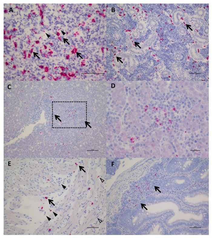
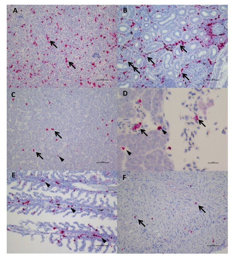
Figure 3. In Situ Hybridization staining of SPAV-1 in Chinook salmon.
The red stain indicates localisation of viral RNA as well as viral transcripts. (A) Spleen: staining mostly localised in the macrophages (arrows) located around the sinusoids, although scattered positive red blood cells (arrowheads) are also present (scale bar 50 µm). (B) Posterior Kidney: the virus appears to be primarily localised in the peritubular capillaries (renal portal vessels) and macrophages (arrows) (scale bar 100 µm). (C) Liver: nodules of inflammation are mainly concentrated in a highly marked area. (scale bar 100 µm), dashed rectangle is enlarged in (D) showing lymphocytes and macrophages in the inflammatory nodule (several of which are positive for the virus). (scale bar 20 µm). (E) Heart: positive macrophages (arrows) are present between the fibres of the spongy myocardium, along with several positive red blood cells (arrowhead) and endothelial cells (open arrowheads). (scale bar 100 µm). (F) Intestine: staining for SPAV-1 is primarily localised to the gut-associated lymphoid tissue (arrows). (scale bar 100 µm).


