. 2019 Aug 21;8:e46112. doi: 10.7554/eLife.46112

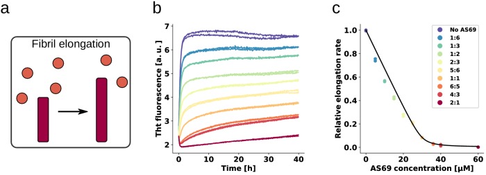
Figure 4. AS69 inhibits α-synuclein fibril elongation.

(a) Schematic representations of fibril elongation. (b) Change in ThT fluorescence when a 30 μM solution of monomeric α-synuclein was incubated in the presence of 5 μM pre-formed fibrils under quiescent conditions with increasing concentrations of AS69. (c) Relative rates of fibril elongation with increasing concentrations of AS69. The solid line corresponds to a prediction based on the affinity of AS69 for monomeric α-synuclein (240 nM, Figure 1b [Mirecka et al., 2014], see Appendix 1 for details).

Figure 4.

Figure 4—figure supplement 1. Characterisation of α-synuclein fibrils formed in the presence and absence of AS69 by AFM.

Figure 4—figure supplement 1.

AFM images of 30 μM monomeric α-synuclein incubated with 5 μM pre-formed fibrils, (a) in the absence or (b) presence of 3 μM AS69, or (c) 30 μM AS69 in 20 mM phosphate buffer at pH 6.5 under quiescent conditions at 37°C for 24 h.
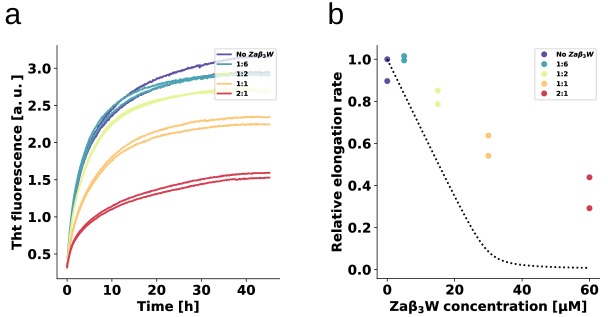
Figure 4—figure supplement 2. Binding specificity determines the inhibitory activity.

Figure 4—figure supplement 2.

The fibril elongation assay was repeated for ZAβ3W, a binding protein for amyloid-β peptide (Grüning et al., 2013) that is a significantly weaker α-synuclein binder than AS69. The absence of detectable heat of binding in ITC allows only an estimate to be made for a minimal Kd of the ZAβ3W-α-synuclein interaction in the order of 10 μM (Mirecka et al., 2014). (a) Change in ThT fluorescence when a 30 μM solution of monomeric α-synuclein was incubated in the presence of 5 μM pre-formed fibrils under quiescent conditions with increasing concentrations of ZAβ3W. (b) Relative rates of fibril elongation with increasing concentrations of ZAβ3W. For comparison to AS69, the dotted line corresponds to the solid line in Figure 4c.