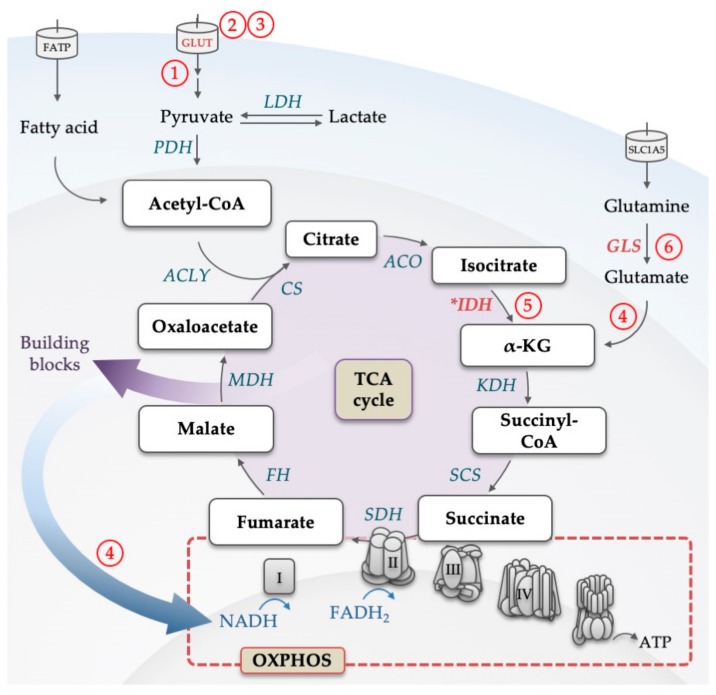
Figure 3.
Illustration of the backbone of the tricarboxylic acid (TCA) cycle. Glucose-derived pyruvate and fatty acids are converted to acetyl-CoA, which feeds the TCA cycle. For each glucose molecule metabolized into the cycle, six NADH, two FADH2 and two ATP (or two GTP) are being produced. Through the conversion to α-KG, also glutamine can replenish the TCA cycle. Products of the cycle are then utilized in the electron transport chain to produce ATP by the oxidative phosphorylation process. Electrons from NADH and FADH2 flow through complexes I, II, III, and IV, which results in a proton gradient across the inner mitochondrial membrane. This in turn provides the energy required to drive ATP synthesis via ATP synthase. Intermediates of the TCA cycle are used as building blocks in other metabolic pathways. Enzymes that are dysregulated in AML are marked in red, mutated are marked with an asterisk. Abbreviations: α-KG, alpha ketoglutarate; ACLY, adenosine triphosphate citrate lyase; ACO, aconitase; ATP, adenosine triphosphate; CoA, coenzyme A; CS, citrate synthase; FADH2, reduced flavin adenine dinucleotide; FATP, fatty acid transporter; FH, fumarate hydratase; GLUT, glucose transporter; GLS, glutaminase; IDH, isocitrate dehydrogenase; KDH, α-KG dehydrogenase; LDH, lactate dehydrogenase; MDH, malate dehydrogenase; NADH, nicotinamide adenine dinucleotide; OXPHOS, oxidative phosphorylation; PDH, pyruvate dehydrogenase; SCS, succinyl-CoA synthase; SDH, succinate dehydrogenase; SLC, solute carrier family; TCA, tricarboxylic acid. Circled numbers refer to metabolic response determinants in AML that are discussed in the Conclusion section.

