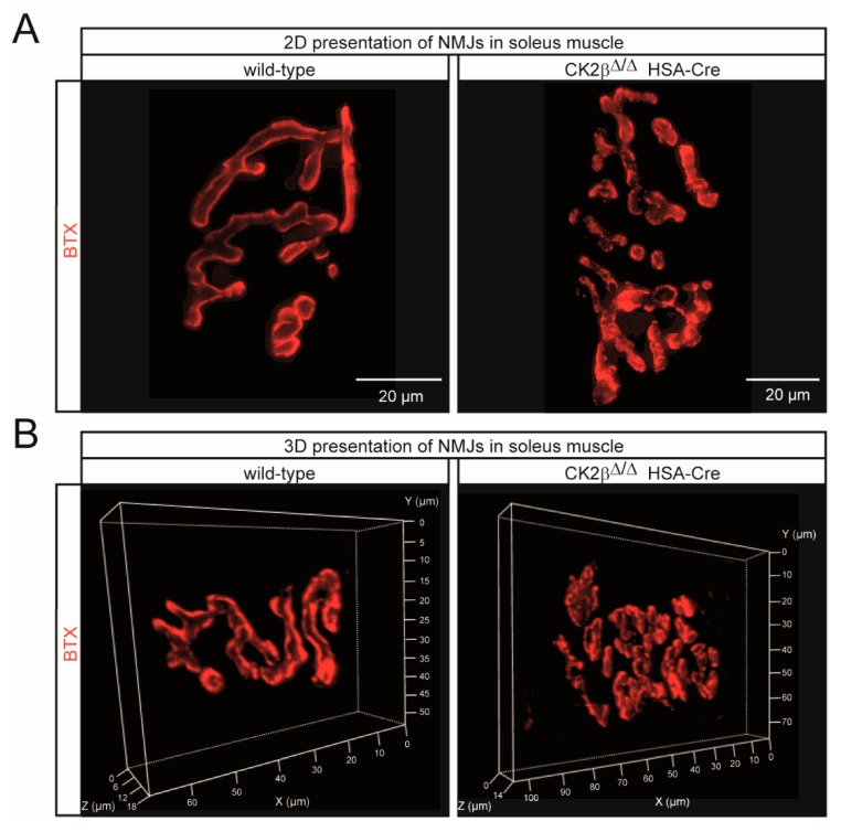
Figure 5.
Conspicuous morphological alterations were visible at the NMJs of CK2β-deficient skeletal muscles. (A, B) Representative 2D (A) and 3D (B) images of whole-mount stainings of wild-type and CK2β-deficient soleus muscle after BTX staining (red) showing the presence of irregular and fragmented NMJs in CK2β-deficient muscles. Scale bar, 20 μm.

