Figure 6.
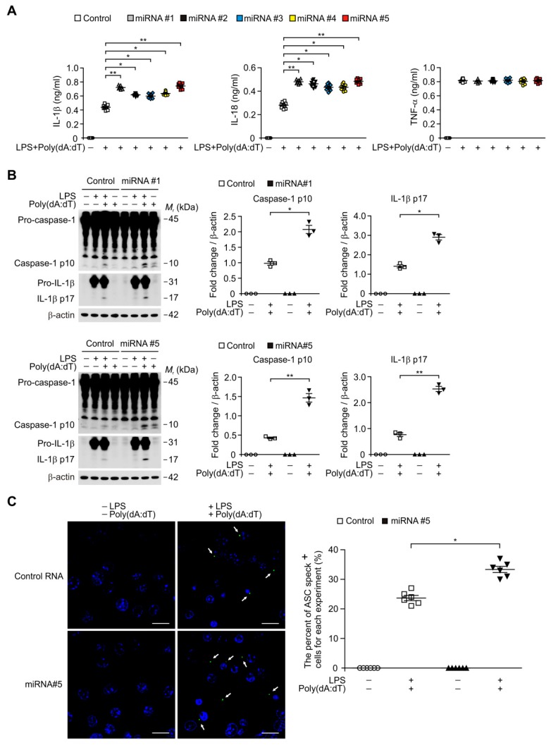
MicroRNA (miRNA) promotes AIM2 inflammasome activation by ASC speck formation in macrophages. (A) Quantification of IL-1β (left), IL-18 (middle), and TNF-α (right) secretion from WT BMDMs transduced with five independent miRNA (miRNA#1, miRNA#2, miRNA#3, miRNA#4 and miRNA#5), or with a control RNA (Control), and stimulated with LPS and poly(dA:dT) (n = 9 mice per group). (B) Representative immunoblot analysis for caspase-1 and IL-1β (left) and densitometry quantification of caspase-1 p10 and IL-1β p17 levels (normalized to levels of β-actin) (right) from WT BMDMs transduced with two independent miRNA (miRNA#1 and miRNA#5), or with a control RNA (Control), and stimulated with LPS and poly(dA:dT). For immunoblots, β-actin was used as loading control. (n = 3 mice per group). (C) Representative immunofluorescence images (total 100 cells in 10 individual images per group) (left) and quantification (right) of ASC speck formation (white arrows; the number of ASC speck-positive cells in 10 individual images per group) in WT BMDMs transduced with miRNA (miRNA#5), or with a control RNA (Control), and stimulated with LPS and poly(dA:dT) (n = 6 mice per group). Scale bars, 20 μm. Data are mean ± SEM. ** p <0.01, * p <0.05; by Student’s two-tailed t-test or ANOVA.

