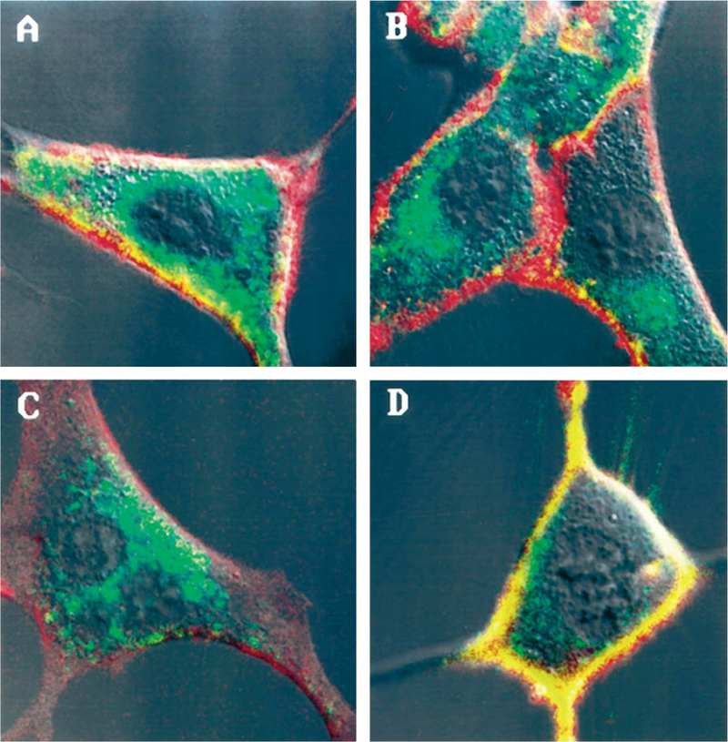
FIG. 4. Internalization of fluorescent Rhodamine Green-conjugated CCK-8 by NIH/3T3 cells stably expressing WT or mutant CCK receptors assessed by confocal laser scanning microscopy.

Shown are representative confocal images of NIH/3T3 cells stably expressing the WT CCKAR (A), the CCKAR ΔS/T (B), the WT CCKBR (C), or the CCKBR ΔS/T (D) after a 15-min incubation with Rhodamine Green-conjugated CCK-8. Cell surfaces labeled with Rhodamine B-conjugated concanavalin A are shown in red. Internalized Rhodamine Green-conjugated CCK-8 is shown in green. Colocalization of concanavalin A and Rhodamine Green-conjugated CCK-8 on the cell surface appears as yellow.
