Figure 3.
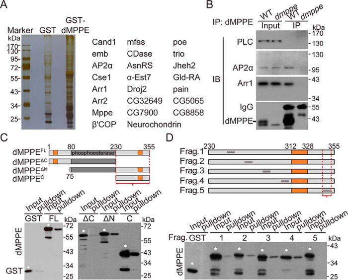
dMPPE directly interacts with Arr1. A, pulldown assay followed by mass spectrum analysis revealed proteins associated with dMPPE. The major dMPPE-associated proteins are listed in the right panel. B, co-immunoprecipitation of dMPPE with Arr1 and AP2α in vivo. Fly head extracts from either WT or dmppe mutants were immunoprecipitated with anti-dMPPE antibody. The precipitates, as well as 4% of the head extract, were subjected to Western blotting with antibody against dMPPE, Arr1, AP2α, and PLC (negative control). IP, immunoprecipitated. C, pulldown assays were performed to map the C-terminal region (bracket) as an Arr1-binding site of dMPPE. MBP-Arr1 fusion proteins were coupled to amylase resin for the pulldown assay. The pulldown samples, as well as a portion (10% of the input for pulldown) of the purified GST-fused dMPPE fragments, were loaded for Western blot analysis. Various GST-dMPPE fusion fragments used for the pulldown assay are indicated by white stars. The other low molecular bands in each lane are degraded products. Upper panel, encoded regions of dMPPE fragments; the transmembrane domains are marked in orange. Bottom panel, Western blots of pulldown assays. D, alanine scanning mutagenesis mapped residues 344–352 (bracket) as an Arr1-binding site of dMPPE. MBP-Arr1 fusion proteins were coupled to amylase resin and incubated with various purified GST-dMPPE fusion fragments. The GST-dMPPE fusion fragments used for the pulldown assays are indicated with white stars. The other low molecular bands in each lane are degraded products. Upper panel, schematic depiction of protein sequences substituted with alanines in the mutant dMPPE; the transmembrane domain is marked in tan. Bottom panel, Western blots of pulldown assays.

