Figure 3.
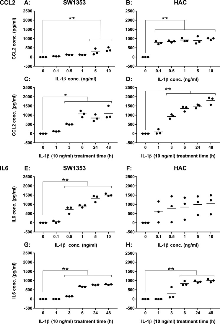
Cytokine secretion levels following exposure to IL-1β. A, CCL2 levels in SW1353 medium following 24-h exposure to IL-1β at concentrations ranging from 0 to 10 ng/ml. B, CCL2 levels in HAC (M70, M76, and M86) medium following 24-h exposure to IL-1β at concentrations ranging from 0 to 10 ng/ml. C, CCL2 levels in SW1353 medium following 10 ng/ml IL-1β treatment for 0–48 h. D, CCL2 levels in HAC (M69, F80, and F86) medium following 10 ng/ml IL-1β treatment for 0–48 h. E, IL-6 levels in SW1353 medium following 24-h exposure to IL-1β at concentrations ranging from 0 to 10 ng/ml. F, IL-6 levels in HAC (M70, M76, and M86) medium following 24-h exposure to IL-1β at concentrations ranging from 0 to 10 ng/ml. G, IL-6 levels in SW1353 medium following 10 ng/ml IL-1β treatment for 0–48 h. H, IL-6 levels in HAC (M69, F80, and F86) medium following 10 ng/ml IL-1β treatment for 0–48 h. Data were considered significant if p = <0.05 (*) and p = <0.01 (**) following Dunnett's multiple comparison tests.

