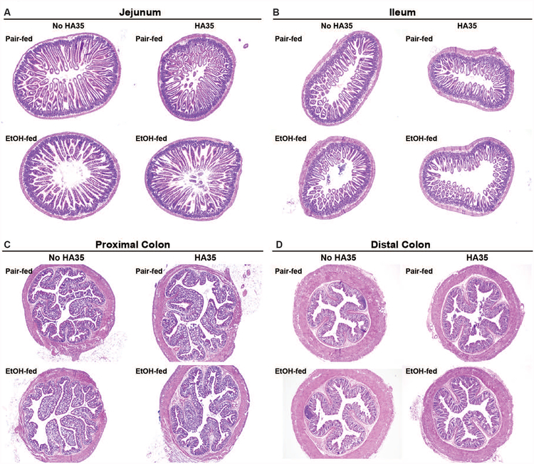
Figure 1: Histology of the intestine in response to short-term ethanol feeding and treatment with HA35.
C57BL/6J mice were allowed free access to an ethanol containing diet (2 days at 11% of calories as ethanol then 2 days at 32% of calories as ethanol) or pair-fed an isocaloric control diet. Mice were provided with 15mg/kg body weight HA35 or an equivalent volume of saline by gavage during the last three days of the short-term ethanol feeding protocol. Paraffin-embedded sections of formalin-fixed (A) jejunum, (B) ileum, (C) proximal colon and (D) distal colon were de-paraffinized and stained with hematoxylin and eosin. Images acquired at 4X magnification. Images are representative of duplicate images captured from n=4 pair-fed or n=6 ethanol-fed mice in each treatment group.

