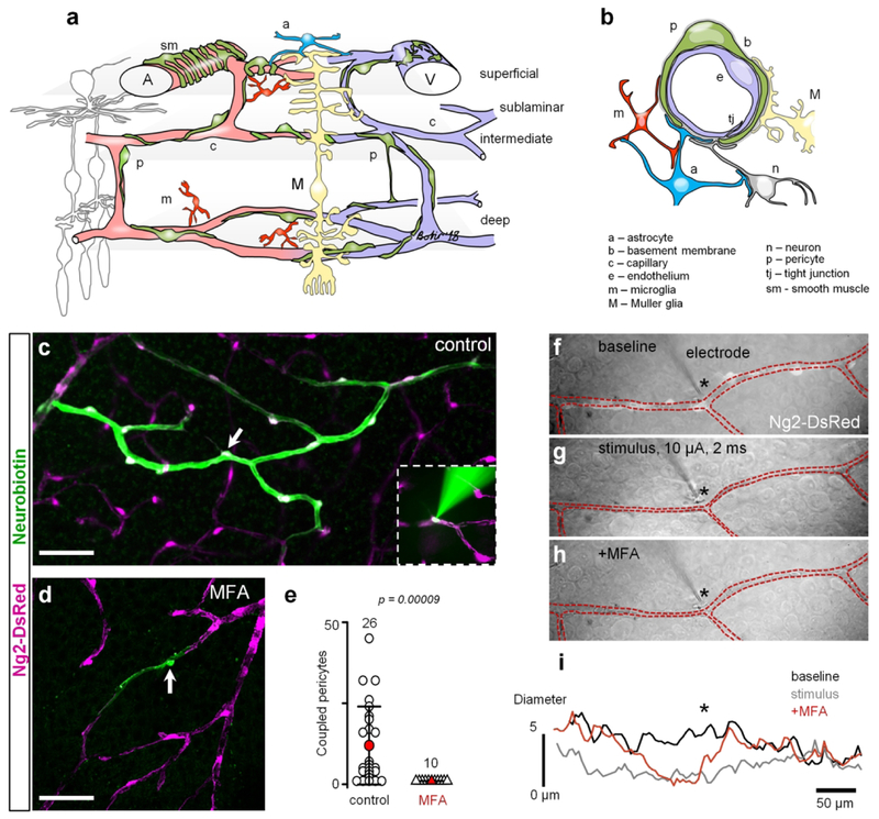
Figure 1. GJs mediate intercellular communication and propagation of vasomotor response.
(a) Specialized domains of the vascular tree. (b) Cross-section of the neurovascular unit. (c) In the living retina whole mount of NG2-DsRed mice, in which all contractile vascular cells express DsRed (magenta), Neurobiotin (green) injected into a single pericyte (arrow) spread to neighboring vascular cells through GJs. Inset shows electroporation procedure. (d) In the presence of pan-GJ blocker meclofenamenic acid (MFA, 50 μM), Neurobiotin spread was limited to the injected pericyte (arrow). (e) Quantification of the Neurobiotin spread in control and in the presence of MFA shows that under normal conditions, vascular cells are extensively coupled through GJs (data are shown as average ± SD; control: 11.96 ± 12, 26 samples, 15 mice; MFA: 1.0 ± 0.01, 10 samples, 8 mice; T-test, p = 0.00009). (f–h) Under physiological conditions, stimulation of an individual pericyte (identified based on NG2-DsRed labeling, white cells along blood vessel in the top panel) led to constriction of the capillary and to spreading of the constriction to the neighboring regions beyond the processes of the simulated pericyte. In the presence of MFA, stimulation of a pericyte led to vasoconstriction underneath the stimulated pericyte, but did not propagate to the neighboring regions. (i) Quantification of the vasoconstriction and propagation of the vasomotor response determined that GJs mediate propagation of the signal along the capillary and they were not involved in vasoconstriction of the stimulated pericyte. Scale bar 50 μm.

