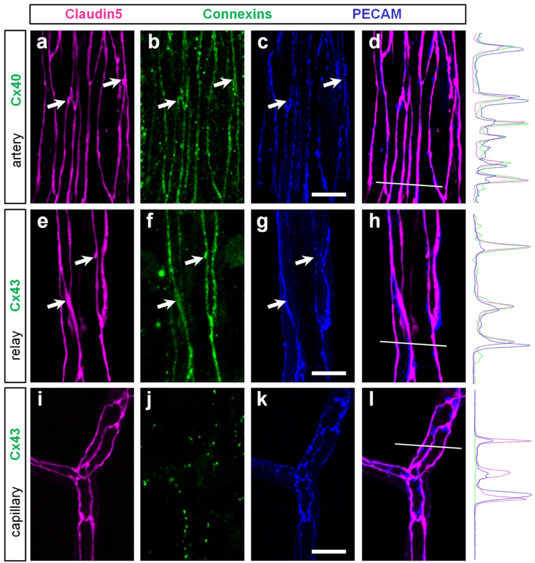
Figure 10. In the arteries and relays gap junctions of endothelial cells strictly colocalize with tight junctions.
(a–d) In the artery, Cx40 (green) was strictly colocalized with tight junctions (magenta, arrows) and less precisely with adherens proteins (blue). In a merged image of claudin5 and PECAM (d), the colocalization was not precise. White line highlights the region used for fluorescent intensity profile. Note that claudin5 and Cx40 profiles closely match and differ from the PECAM profile. (e–h) In the vascular relay, Cx43-positive strings were colocalized with tight junctions including strings and small varicosities (arrows). (i–l) In the capillary, Cx43 was not detected along tight junctions. Scale bar 10 μm.

