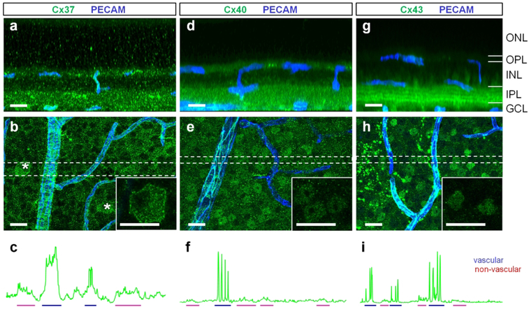
Figure 3. Cx37, Cx40, and Cx43 are expressed in retinal neurons at much lower levels than in the vasculature.
(a) Cx37 expression shown in a vertical view, created by rotation of a z-stack from the corresponding retinal whole mount region in (b). (b) Confocal projection of a retinal whole mount at the GCL. Some ganglion cells expressed Cx37 at their cell membrane (asterisks, insert). (c) Blood vessels had higher expression of Cx37 (blue) than ganglion cells (red). (d–e) Cx40 was weakly expressed in numerous nuclei of inner retinal neurons. High expression of Cx40 was detected in blood vessels (f, blue). (g–i) Cx43 was expressed in nuclei (insert) and processes of inner retinal neurons (g, four bright bands in the inner plexiform layer). The expression in neurons was lower than in blood vessels (i). Scale bar 20 μm. ONL – outer nuclear layer, OPL – outer plexiform layer, INL – inner nuclear layer, IPL – inner plexiform layer, GCL – ganglion cell layer.

