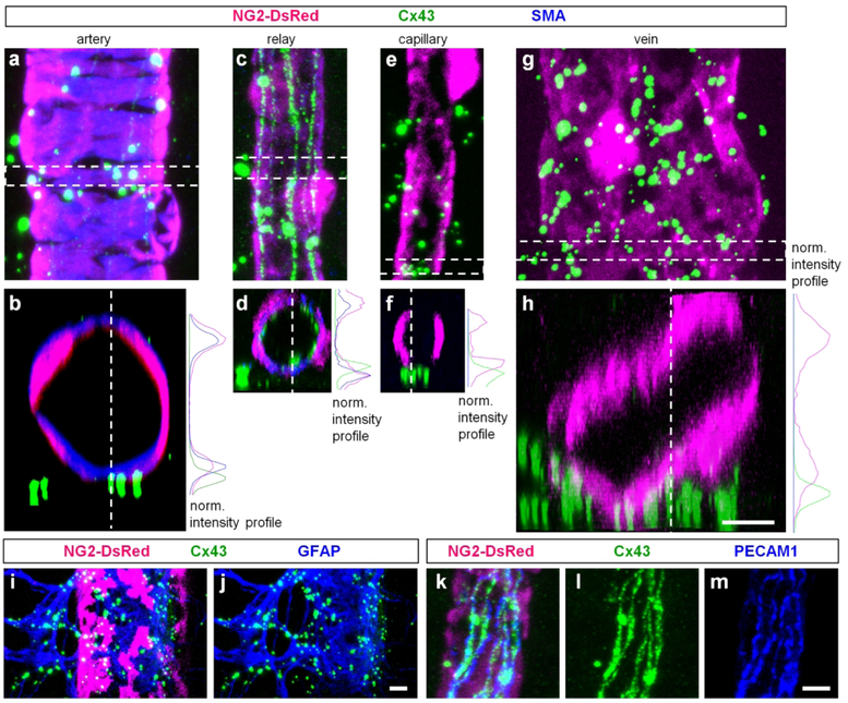
Figure 5. Cx43 is primarily localized to astrocytes and vascular cells.
(a) Around an artery, majority of the large puncta was not directly associated with vascular cells. (b) A vertical view from the highlighted area in (a) was obtained by 900 rotation of the z-stack. On the right, fluorescence intensity profile for the three labeling from (b). (c–d) In the relay, large puncta were also not directly located on the blood vessels contrarily to the string-like structures which were enclosed by the mural cells (d panel and the intensity profile). (e–f) Around the finest capillaries, Cx43-puncta were mostly outside of the blood vessels. (h) In veins, putative labeling was not directly associated with mural cells. (i–j) Bright Cx43 puncta around blood vessels were localized to astroglia labeled for glial fibrillary acidic protein (GFAP). (k–m) Cx43-positive strings were associated with endothelial cells labeled for platelet endothelial cell adhesion molecule-1 (PECAM-1). Scale bar 5 μm.

