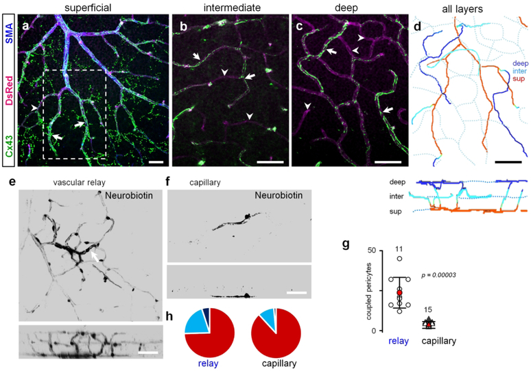
Figure 6. Cx43 promotes vascular coupling between the capillaries and the artery.
(a-c) The relay zones were identified based on the staining against smooth muscle actin (SMA). The string-like Cx43-positive structures started at the primary artery and continued uninterrupted beyond SMA-labeling into selected capillaries (arrows). Some capillaries had much weaker or no string-like expression (arrowheads). The strings from the superficial layer in (a) extended into intermediate (b) and deep (c) vascular layer capillaries using the shortest route from the artery. Scale bar 50 μm. (d) In the projection of all layers, string-bearing selected capillaries created a specialized vascular domain – vascular relay - spanning all three layers. The bottom panel shows a vertical view. Scale bar 50 μm. (e) Neurobiotin injected into a pericyte (arrow) in relay area spread throughout the large area. (f) Neurobiotin injected into the area outside the relay was restricted to a few pericytes. (g) Quantification shows that under normal conditions cells are extensively coupled through GJs in vascular relay (data are shown as average ± SD; standard: 3.3 ± 2.1, 15 samples, 8 mice; sensor: 23.7 ± 9.6, 11 samples, 6 mice; T-test, p = 0.00003). (h) Distribution of Cx43-positive strings across vascular layers shows that in the vascular relay region Neurobiotin spread farther into intermediate and deep vascular layers.

