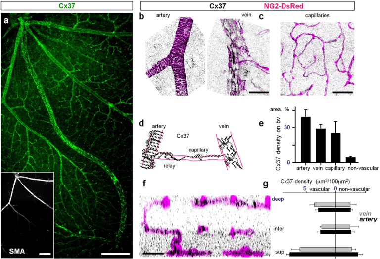
Figure 7. Cx37 is expressed in mural cells.
(a) Cx37 was located on all types of blood vessels in a retina whole mount. Insert identifies arteries based on the presence of smooth muscle actin labeling (SMA). Scale bar 200 μm. (b–c) In high magnification contractile cells identified by DsRed labeling express Cx37. (d) Schematic presentation of Cx37 distribution on retinal vasculature. (e) Cx37 density was high on all types of vasculature. (f) Cx37 distribution in a vertical retinal view shows punctate labeling in the inner plexiform layer. (g) Density of Cx37-labeled structures in all vascular layers. Scale bar 50 μm.

