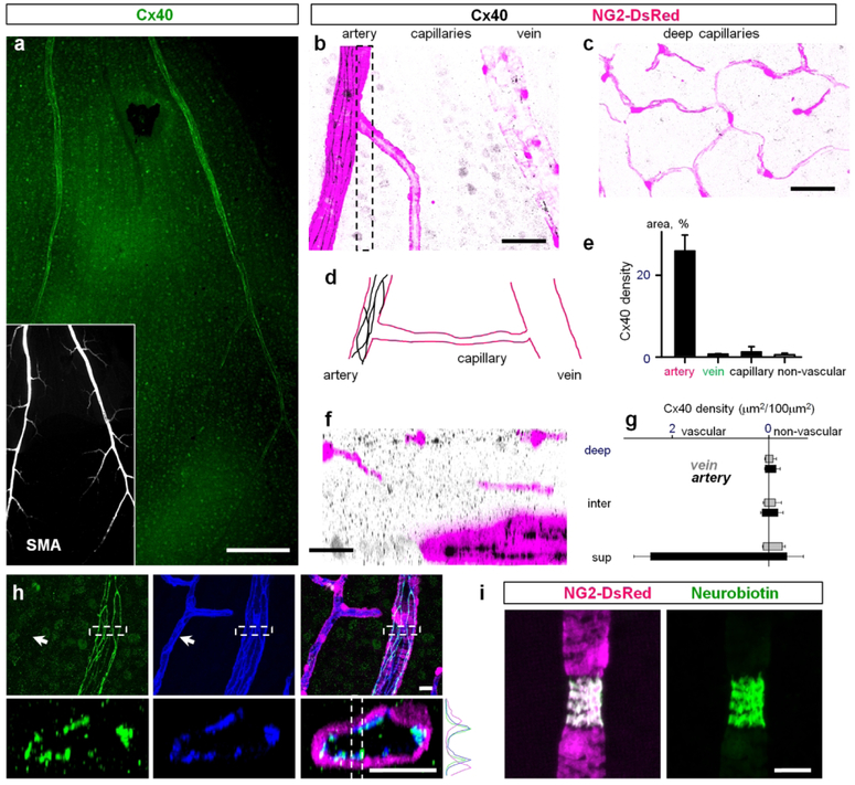
Figure 9. Cx40 was primarily localized to endothelial cells of the primary arteries.
(a) Cx40 was associated with primary arteries and some neurons in the ganglion cell layer of the retina whole mount. Insert shows primary arteries labeled for smooth muscle actin (SMA). Scale bar 200 μm. (b) In a high magnification of the superficial vascular layer Cx40 was localized directly to arteries. Scale bar 50 μm. (c) Cx40 was not detected in neither intermediate layer nor deep layer capillaries. Scale bar 50 μm. (d) Schematic presentation of Cx40 distribution in the superficial layer. (e) Quantification of Cx40 distribution on blood vessels of the superficial layer. (f) Vertical view of the confocal z-stack through the area highlighted in (b). Scale bar 25 μm. (g) Density of Cx40 in vascular layers. (h–j) Cx40 was colocalized on endothelial cells of the primary arteries but not on the capillaries (arrow). (i) Neurobiotin injected into a smooth muscle cell did not propagate to neighboring vascular cells. Please note that the targeted SMA-positive cell constricted during Neurobiotin injection. Scale bar 25 μm.

