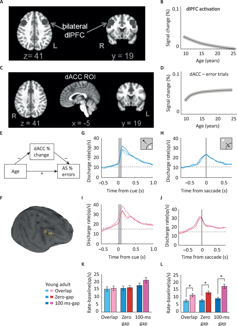
Figure 2. Maturation of response inhibition in imaging and physiological studies.
A. image of human brain indicating areas in the dorsolateral prefrontal cortex (dlPFC) that undergo changes in AS task activation during adolescence. B. Schematic diagram of mean growth curve indicating dlPFC signal changes with age. Of executive control regions, only the right dlPFC demonstrates developmental changes in activation. C. Image of human brain indicating areas in the dorsal Anterior Cingulate Cortex (dACC) that undergo changes in activation during adolescence. D. Schematic diagram of signal change in dACC with age. dACC activation is associated with better overall task performance, as indicated by lower AS error rates. E. Signal change in the dACC during corrected error trials mediates the effect of age on AS error rates. F. MRI image of an adolescent monkey with areas 8 and 46 of the dlPFC indicated. Arcuate Sulcus (AS) and Principal Sulcus (PS) are also shown in the figure. G-J. Mean firing rate in the young (G-H) and adult (I-J) stage for monkeys, in three variants of the antisaccade task (overlap, zero-gap, gap). Responses are shown for stimulus in the receptive field (G,I) and for saccade in the receptive field (H,J). K-L. Histograms average firing rates relative to baseline in a 200 ms window, for the stimulus in the receptive field (K) and saccade in the receptive field condition (L). No decrease was observed in activation elicited by the visual stimulus in adulthood (G,I,K). A significant increase in activity was present in adulthood for the representation of the goal of the saccade (H,J,L). Panels A-E adapted with permission from [6]; Panels F-L from [32].

