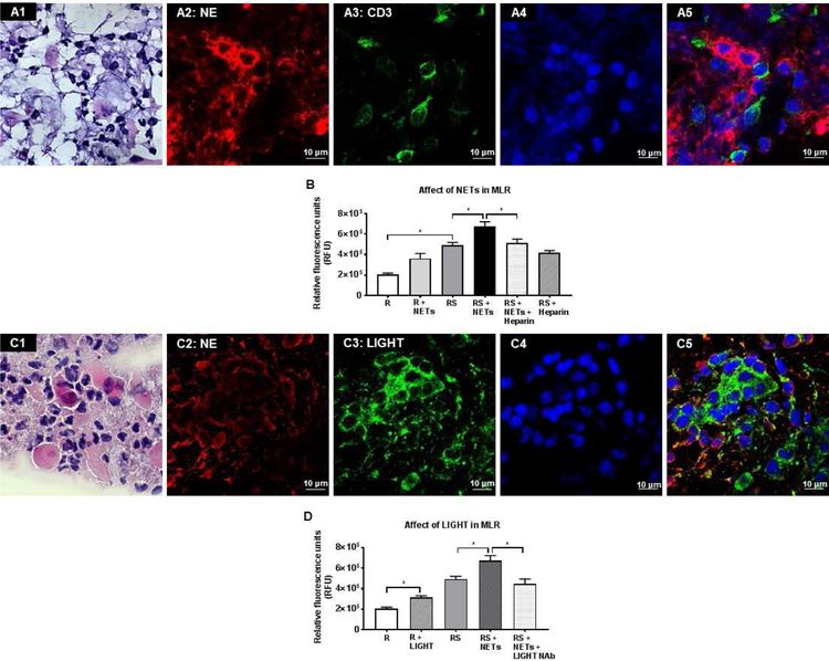
Figure 8. Pathological effect of NETs on immune cell proliferation.
(A1): H&E staining of mucocellular aggregates (MCA) showed numerous neutrophils, surface epithelial cells and mononuclear cells. (A2-A5): Confocal immunofluorescent staining images of MCA showing the presence of neutrophil elastase (NE) (A2, red), CD3 positive T cells (A3, green), DAPI nuclear staining (A4, blue) and merged image (A5). (B): Graph shows NET-induced T cell proliferation in mixed lymphocyte reaction (MLR). NETs promote proliferation of MLR. Heparin inhibits MLR proliferation. (C1): H&E staining of mucocellular aggregates (MCA) showed numerous neutrophils, surface epithelial cells and mononuclear cells. (C2-C5): Confocal immunofluorescent staining images of MCA showing the presence of neutrophil elastase (NE) (C2, red), LIGHT protein (C3, green), DAPI nuclear staining (C4, blue) and merged image (C5). (D): Graph shows effect of human recombinant LIGHT/TNFSF14 protein induced on T cell proliferation and in MLR. LIGHT/TNFSF14 induces T cell proliferation. LIGHT/TNFSF14 neutralizing antibodies inhibit NET-induced MLR proliferation.

