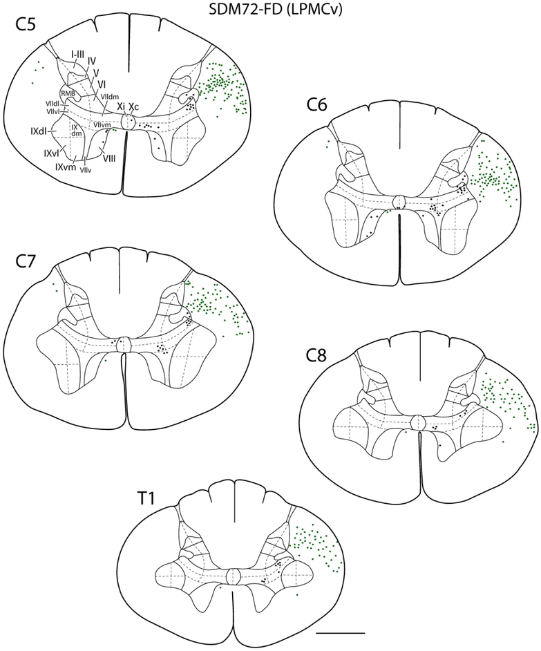
Figure 5.
Line drawings of representative transverse sections through spinal levels C5-T1 from LPMCv case SDM72-FD depicting the locations labeled white matter axons (green dots) and locations of labeled gray matter axon terminals (black dots). Spinal levels are depicted on the top left of each transverse section. Roman numerals in section C5 denote Rexed’s laminae and their respective anatomical subsectors used for quantitative stereological analysis of terminal bouton numbers. For orientation dorsal is located on the top of each section and ventral at the bottom. Note the significantly lighter distribution of spinal terminals compared to the LPMCd cases shown in Figure 4. Abbreviations, dm, dorsomedial; dl, dorsolateral; v, ventral; vl, ventrolateral; vm, ventromedial. Scale bar = 2mm.

