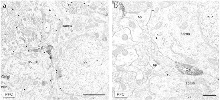
Figure 2: CB1-containing axon terminals positioned between two neuronal soma.
(a) A large CB1-labeled axon terminal (CB1) is sandwiched between two mGluR5-labeled soma in layer III of the mPFC. Another lightly labeled, irregularly shaped CB1-containing terminal contacts the upper soma at a point of dendritic branching. (b) An axon terminal containing CB1 immunoreactivity is positioned between two tightly apposed neuronal soma, also in layer III of the mPFC. Scale bar (a) = 2 μm, scale bar (b) = 500 nm, CB1 = CB1-containing axon terminal, Golgi = Golgi apparatus, nuc = nucleus, sp = dendritic spine.

