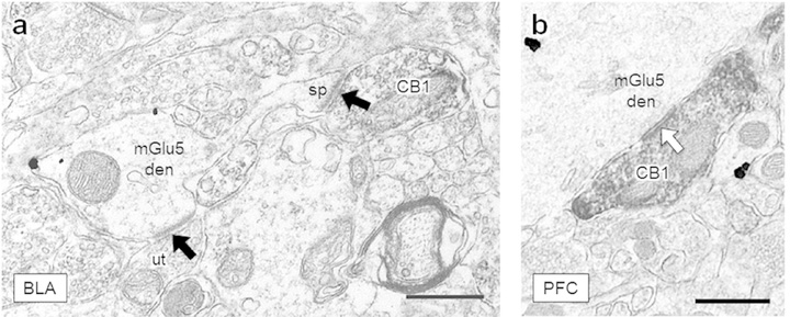
Figure 3: CB1-containing axon terminals form synapses in the BLA and PFC.
(a) A CB1-labeled axon terminal forms an asymmetric excitatory-type synapse (black arrow) with a spine protruding from a nearby mGlu5-containing dendrite in the BLA. An unlabeled terminal (ut) forms an excitatory synapse onto the dendritic shaft. (b) An axon terminal containing CB1 immunoreactivity forms a symmetric inhibitory-type specialization (white arrow) with a large dendrite. Scale bars = 500 nm, CB1 = CB1-containing terminal, mGlu5 den = dendrite containing mGlu5 immunogold, ut = unlabeled terminal, sp = dendritic spine.

