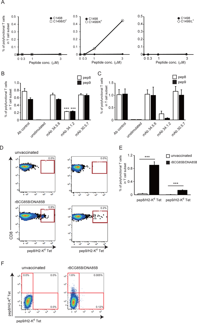
Figure 6. H2-Kd alloantigen specificity for induction of the epitope peptide-stimulated polyfunctional T cells.

(A) C1498 cells transfected with H2-Dd, H2-Kd or H2-Ld were incubated with pep9 peptide and used as stimulator cells for polyfunctional T cell induction with various ratio of effector and stimulator cells; 0, 0.3, 1 and 3. Splenocytes from BALB/c mice immunized with rBCG-Mkan85B/DNA-Mkan85B were incubated in the presence of medium alone or the stimulator cells for 6 h and the polyfunctional T cell induction was analyzed by flow cytometry. (B and C) MHC-I alloantigen specificity of pep8 and pep9. Spleen cells from rBCG-Mkan85B/DNA-Mkan85B immunized BALB/c (B) and CB6F1 (C) mice were incubated with mAb 34.5.8 (anti-H-2Dd antibody), mAb 34.1.2 (anti-H2-KdDd antibody) or mAb 30.5.7 (anti-H2-Ld antibody), followed by stimulation with the pep8 or pep9 for polyfunctional T cell assays. (D) Representative fluorogram of reactivity of pep8/H-2Kd and pep9/H-2Kd tetramer-positive CD8+ T cells from rBCG-Mkan85B/DNA-Mkan85B immunized BALB/c mice, with pep8/H-2Kd (left panel) or pep9/H-2Kd (right panel) on x-axis and CD8 on y-axis. Control refers to immunization with vector alone. Spleen cells from BALB/c mice were analyzed for reactivity with the pep8/H-2Kd tetramer conjugated to allophycocyanin (APC) or with the pep9 /H-2Kd tetramer conjugated to phycoerythrin (PE). (E) pep8/H-2Kd and pep9 /H-2Kd tetramer-positive CD8+ cell responses in the mice immunized with rBCG-Mkan85B/DNA-Mkan85B (closed bar) or control (open bar). Data represent three independent experiments with five mice per group. ***, p < 0.001; one-way ANOVA test. Error bars represent SEM. (F) Representative fluorogram of double staining with the two tetramers labelled with different fluorochromes with pep9/H-2Kd on x-axis and pep8/H-2Kd on y-axis. Percentage of positive cells is indicated in each quadrant.
