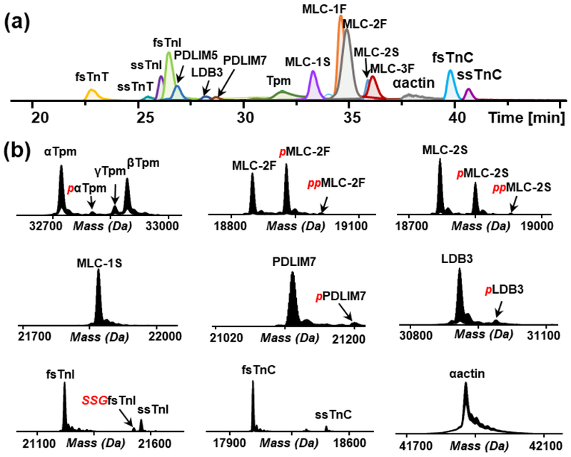
Figure 1.
LC/MS analysis of myofilament proteins. (a) LC/MS extracted ion chromatograms of myofilament proteins from rhesus macaque vastus lateralis (VL) tissue extract. (b) Deconvoluted mass spectra showing proteoforms of selected myofilament proteins, Tpm, MLC-2F, MLC-2S, MLC-1S, PDLIM7, LDB3, TnI, TnC and αactin. Red italic p, phosphorylation; Red italic SSG, S-gluthathionylation.

