Fig. 5.
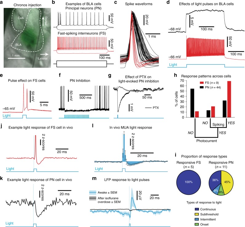
In vitro validation of optogenetic strategy. a Chronos injection (green) in the BLA. b Example recordings of BLA principal neurons (PN, black) and fast-spiking interneurons (FS, red). c Spike waveforms (n = 53 cells, 10 rats). d Example responses to a 500-ms light pulse (blue). e FS responses to 2-ms light pulses (blue). f PN inhibition by a light-pulse train (blue). g Effect of picrotoxin (PTX-100 μM, gray) on light-evoked inhibition of PN (black). h Cell response patterns (PN-black, FS-red). i Proportion of in vitro spiking at 50 Hz light stimulation (top) along with prevalent response types (bottom). j Example response of FS cell to light pulses in vivo. k Same for example PN cell. l In vivo multiunit activity (MUA) response to 2-ms light pulses (n = 6 sessions, 3 rats). Error bars: SEM. m Light-evoked LFP potential (average, blue ± SEM, shading) by 2-ms pulses is abolished by isoflurane overdose (black, n = 5 rats, Mann Whitney U(4) = 40, p = 0.0079)

