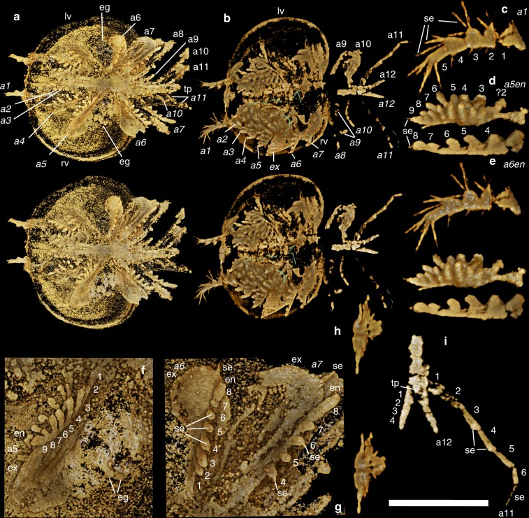
Fig. 1.
Kunmingella douvillei (Mansuy, 1912): a, f, g (YKLP 16235); b–e, h, i (YKLP 16233). All views are ventral. a–e, h Stereo-pairs with a 20° tilt. a, b Whole animal views: in a, eggs can be seen mid-valve, nestled in the region of the lobal structure; in b, the green coloured elongate structures appear to be organic but are not part of the bradoriid. c Right appendage 1. d Endopod of right appendage 5 (incomplete proximal part). e Endopod of right appendage 6 (proximal podomeres not shown). f Right appendage 5. g Left mid and some posterior trunk appendages. h Possible neural structures. i Posterior-most part of the trunk and limbs. Scale bar: a 2.19 mm; b 1.65 mm; c 560 µm; d 850 µm; e 610 µm; f 820 µm; g 730 µm.; h 680 µm; i 810 µm. a1–a12 1st to 12th appendage (italic signifies a right-side appendage), eg egg(s), en endopod, ex exopod, se seta(e), tp tailpiece, numbers 1–9 refer to the podomeres and/or endites, from distal to proximal

