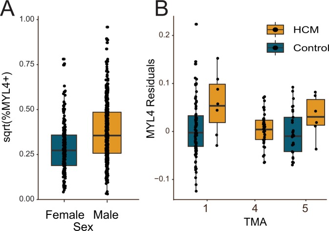
Figure 2.
MYL4+mosaicism by gender and disease. (A) Male subjects had more MYL4+ cells, independent of disease state (p = 8.66e-15). (B) In left ventricular segments, MYL4+ cells were increased in HCM compared to control tissues (TMAs 1&5). There were no control septal tissues to compare to the HCM cases on TMA4. The MYL4 residual is of a regression of sqrt(%MYL4+) cells adjusted for multiple measures per individual and effects of different TMAs, age, and sex.

