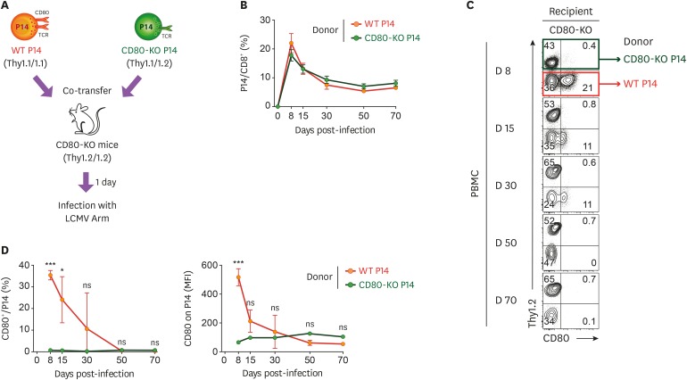
Figure 2. The intrinsic expression of CD80 on Ag-specific CD8+ T cells gradually decreases in the blood. (A) Experimental scheme for investigation of intrinsic expression and extrinsic acquisition of CD80 on Ag-specific CD8+ T cells in the blood. WT P14 CD8+ and CD80-KO P14 CD8+ T cells were adoptively transferred together into CD80-KO recipient mice. One day after the adoptive transfer, mice were intraperitoneally infected with 2×10⁵ PFUs LCMV Arm. (B) Frequency of donor WT and CD80-KO P14 CD8+ T cells among CD8+ T cells obtained from PBMCs of CD80-KO recipient mice at the days indicated post-infection. (C) Expression of CD80 on WT and CD80-KO P14 CD8+ T cells among Thy1.1+ donor cells obtained from PBMCs of CD80-KO mice at the days indicated post-infection. (D) Frequency of CD80-expressing cells and MFI of CD80 among WT and CD80-KO P14 CD8+ T cells obtained from PBMCs of CD80-KO mice at the days indicated post-infection. Data are representative of 3 independent experiments (n=3−4 mice per group). Results represent the mean±SEM and statistical significance was determined by 2-tailed unpaired Student's t-test.
NS, not significant; MFI, mean fluorescence intensity.
*p<0.05; ***p<0.001.

