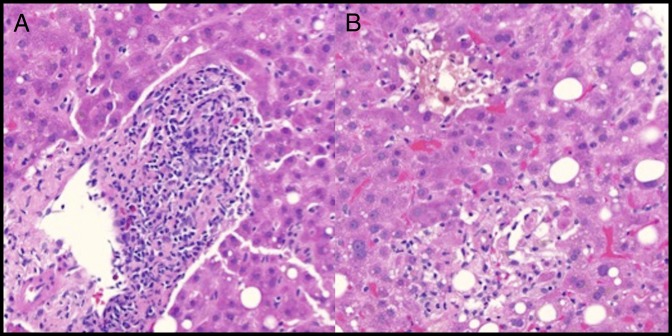
Figure 2.
(A) Hematoxylin and eosin (H&E) stain of the pretransplant liver biopsy showing mixed portal inflammation, including lymphocytes, plasma cells, and eosinophils (400×). Although present, plasma cells are not predominant. The portal vein and artery are readily identifiable, but the duct is heavily infiltrated by inflammatory cells. No significant interface activity. (B) H&E stain of the pretransplant liver biopsy showing marked lobular cholestasis with associated hepatocyte dropout and mild lobular lymphocytic inflammation (400×).

