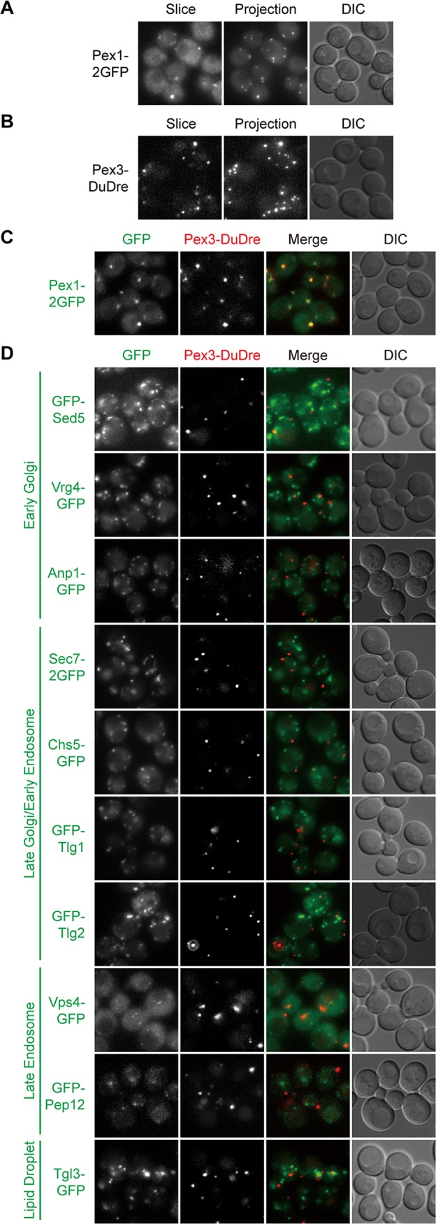
FIG 6.

Green and red markers of peroxisomes. Images were captured and presented as in Fig. 1. (A and B) Green (A) and red (B) markers of peroxisomes. (C) Colocalization between green and red peroxisome markers. (D) Lack of colocalization between green markers of other organelles and the red marker of peroxisomes.
