Fig.1.
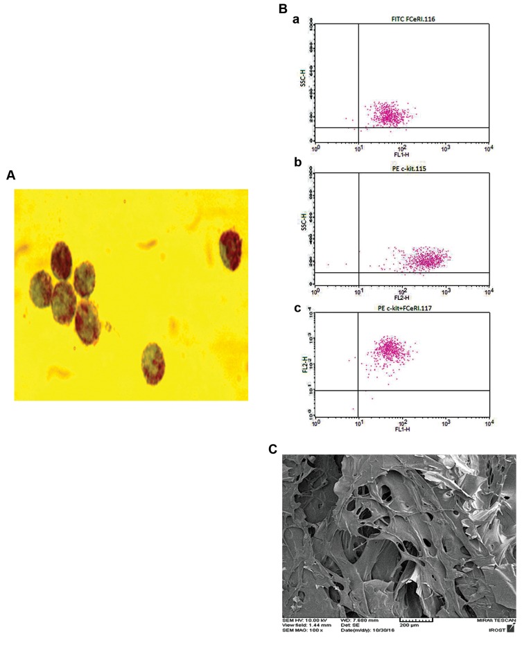
Presentation of the cell characterizations and scaffold microstructure. A. Murine bone marrow mast cells (BMMC) were cultured in pokeweed mitogen-stimulated spleen cell conditioned medium (PWMSCM), 20% (v/v) for 3 weeks and cells were stained with toluidine blue (representative image at ×1000 magnification), B. Representative flow cytometry analysis of BMMC. (a) Cells positive for FC.RI, (b) Cells positive for CD117 (c-kit), and (c) Double positive cells (92%), and C. Representative micrograph of scanning electron microscope to evaluate ultra-structure of porosity of the chitosan scaffold. Images are representative of at least n=3 independent experiments.

